Cattle with normal care becomes a tangible help for a small farm. Unfortunately, cows and calves sometimes get sick, which immediately affects productivity or growth. The "secretive" nature of many diseases and the absence of overt symptoms are added to the complexity.
Consider one of the most massive ailments among animals, namely cysticercosis (aka finnoz).
What is it and how dangerous
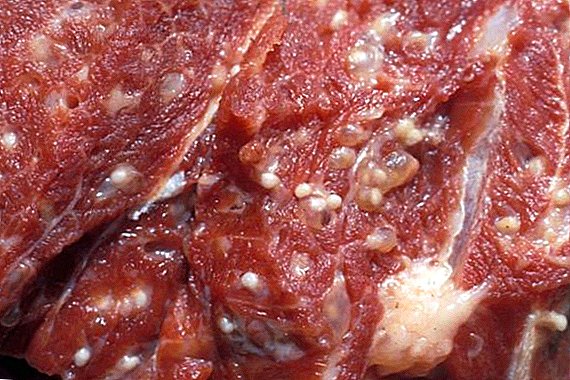
This is a parasitic disease caused by cysticercus - a bull chain at the larva stage. The causative agent is a vial with a scolex (a head with four suckers), filled with turbid liquid. Such a "ball" is covered with villi and differs in rather large sizes (up to 8-9 mm in length and 5-6 in width).
The harmful larva affects the muscles - the muscles of the skeletal group, heart and tongue, as well as internal chewing. The danger lies in the fact that with a weakened immune system may be affected adipose tissue and liver, brain and lungs. 
How is the infection of animals and who is the carrier
Cysticerci can be ingested with water and food in which eggs or mature chains are present. The "catalyst" is gastric juice, softening their shells and accelerating the release of embryos (oncospheres).
Important! Do not forget about elementary hygiene: a fly that has sat down on food will last 4-5 seconds to “throw” a chain of eggs. It is advisable to hide the meat immediately in the refrigerator.They, having barely left the egg, penetrate into the mucous layer of the intestinal tract, blood vessels and interfibrillary parts of the muscles. This helps six strong hooks.
The infection pattern is traditional and simple, but cysticercosis is impossible without human interventionbecause it is in his organism that the pathogen reaches the stage of maturity. Scientifically, people are definitive owners of this parasite, while animals themselves are intermediate.

The larvae enter the human body along with the food (most often the uncooked meat of the affected cattle). The chain may not cause any reactions in people, being inside for many years and sometimes growing up to 10 m.
Among the diseases of cows emit mastitis, ketosis, leukemia, anthrax, brucellosis, dyspepsia, foot and mouth disease, tuberculosis, gastroenteritis.After 2-3 months, the parasite begins to produce eggs that go out with feces. Therefore, the risk group includes animals that live in places where there is no normal drainage, and sewage gets into pastures. Another route of infection is contact with already infected farm workers or stray animals.
Life cycle
Barely appeared oncospheres become full-fledged larvae 5-6 months after their appearance. Faster rates were noted (3.5-4 months), but this is more characteristic of parasites that settle on the walls of the blood arteries.
Did you know? Until 1784, the larvae of these parasites were considered a separate helminth. But Johann Gosier, in the course of his research, found out that oncospheres are nothing more than the “offspring” of bovine tsepny.The peak of activity accounts for 7-10 months of presence: at this time, you can recognize the characteristic symptoms of the disease. After a 10-month "turn" the larvae gradually die off. This process can take another month and a half.

Signs of disease
The cunning of finnoza is that with a low degree of invasion (infection), the state of health of the cattle does not inspire fear - the symptoms are practically not manifested.
But the imposing cysticerci colony will surely “give out” itself, bringing the matter to an acute form. It can last for two weeks - the first 5-6 days the course of the disease is particularly clear, after which the characteristic signs disappear. Among them are:
- a sharp decrease in appetite or a complete rejection of feed;
- increased body temperature;
- the anxiety of animals, they become very agitated;
- frequent diarrhea;
- dry mucous membranes, which also fade;
- “malfunctions” in the heart’s work, frequent dyspnea indicate this; when moving to pasture, usually an active cow can stop many times;
- muscle pain;
- painful reaction to palpation in the net and rennet area.

Important! In a sick calf, the temperature can “catch up” from 39.8 ° C to 41.7 ° C. To feel it, you do not even need a thermometer - the difference is already visible with the usual touch of the palm.Complications such as pruritus, ascites, or blindness are rare. The most dangerous symptom of veterinarians is called a drop in temperature, which can in a day or two can result in the death of an animal. Fortunately, such manifestations are extremely rare.
Pathological changes
If the disease occurs in an acute or chronic form with background complications, the structure of the affected areas is invariably disrupted. When cutting up dead animals, characteristic "traces" of larvae are found on such organs and tissues:
- numerous point hemorrhages;
- the presence of small cysticerci;
- an increase in the mesentery, the lymphatic ligament itself in the section has an unnaturally juicy tint;
- discoloration of skeletal muscles (they turn gray), on which deposits in the form of light gray oncospheres can be found.

Diagnostics
Making an accurate diagnosis is a problem even for experienced veterinarians.
The fact is that some symptoms (for example, loss of appetite or anxiety) are considered indirect and can "lead" to a completely different disease. Yes, and manual techniques like probing with particular accuracy do not differ - of course, the sick animal will respond with a roar, but it will not be possible to localize the center of infection in this way.
Did you know? In Australia, cases of finnozom are recorded quite rarely (both among animals and in humans) - the parasite does not tolerate such a climate. Most carriers are immigrants who come from different continents.More or less a complete picture can give the results of special analyzes. Usually appointed:
- Rnga (indirect hemagglutination). This method is used in the study of blood syrup. The collected material in a volume of 5-7 ml is placed in a sterile tube, and then put in a tablet with red blood cells. If a precipitate forms, it is a sure sign that the parasite is in the body. The reaction of the NGA is considered the most accurate of the used analyzes.
- RLA (latexagglutination). Blood syrovotoka warms up, then add a latex suspension, making several single inclusions of other drugs. After the plates with the collection material are “scrolled” on the joker, a conclusion is made. The presence in the statement marks "++" or "++++" indicates that the larvae are actively developing.
- Intradermal allergy test. The drug tuberculin is injected into the middle part of the neck or the tail-tail fold (calves - into the shoulder blade). The dose depends on the age, the norm for adult livestock is 0.2 ml, while young animals up to one year old need 0.15 ml. In healthy animals, 12–20 hours after the injection, edema appears, which increases in 2–3 days. In the delayed reaction (48 hours), another injection is given, the results of which bring complete clarity.

Important! Unwanted "guests" can be found with careful examination of the tongue and mouth. True, the cow may simply not be given in the hands (this is also a symptom, albeit an indirect one).Often used and post-mortem inspection of carcasses. First, make various incisions of the muscles of the heart, tongue and chewing ligament. For a more accurate result, the lumbar and cervical muscles are cut in the same way.
Larvae become visible when a fluorescent lamp is scanned through a dark room. Parasites are highlighted in red or burgundy. When viewing frozen meat, the effect will be the same, but the cysticercus will die by that time. Boiled meat ceases to "shine" after 1.5 hours of heat treatment.
Is treatment possible
The treatment of cattle diseases such as cysticercosis is complicated due to the specific action of the pathogen.
Prescribing drugs requires considerable experience from the veterinarian. Strong antihistamines are no good here. - their action causes a massive death of parasites, but at the same time, intoxication and inflammation are triggered in the muscle fibers “stuffed” with them.
Praziquantel-Mebendazole and Dronzit formulations are used.. The first drug is added to the feed at the rate of 50 mg / kg. Reception course - 10 days. As for Droncyt, its dose is determined only by the doctor who examined the animal (it is advisable to have the test results on hand). It is not surprising that an important role is assigned to prevention, which can eliminate all these difficulties.
Prevention
It includes a whole range of measures. Required are the following events:
- slaughter cattle only in slaughterhouses that have all the necessary equipment with mandatory inspection of carcasses;
- when more than three larvae are found, control cuts are made;
Did you know? In the 18th century, each artillery regiment necessarily included a horseman and three apprentices. The cavalry regiments were ordered to have already 10 such masters (according to the orders of Peter I, published in 1712).
- as necessary - technical disposal of infected carcasses.
 Of course, you should not forget about sanitary and hygienic factors:
- Arrangement of "latrines" closed.
- Maintain cleanliness in the stall or on the farm.
- Grazing in clean areas, remote from open drainage pits and channels.
- No contact with stray animals.
- For large farms, periodic animal verifications and professional examination of staff are mandatory.
- Finally, thorough processing of meat before eating. Careful roasting or cooking will not give the parasite a chance to walk along the "food-person-animal" chain.
The peculiarities of the content and productivity of different breeds of cows are not identical; the characteristics of Simmental, Dutch, Holstein, Ayrshire, Jersey, Aberdeen-Angus, Red Steppe, Kalmyk, Yaroslavl cows should be studiedNow you know what is the danger of finnoza, how to prevent infection. We hope this information is useful only for reference. Let the economy bring only positive and income!